Gut Dysbiosis and Parkinson’s Disease: 6 Surprising Connections
Explore the critical link between Gut Dysbiosis and Parkinson’s Disease, uncovering how gut health impacts neurological disorders.

In recent years, the intriguing connection between gut dysbiosis and Parkinson's Disease (PD) has garnered significant attention.
This article delves into the complex interplay between the gut-brain axis and its impact on non-motor symptoms in PD, notably pain hypersensitivity.
We explore how alterations in gut microbiota not only influence the severity of PD's motor symptoms but also contribute to the heightened pain sensitivity experienced by patients.
By examining the role of neuro-immunity in this context, we aim to shed light on the underlying mechanisms that link gut health to neurodegenerative processes, offering new perspectives on managing and understanding PD.
In This Article:
Key Findings
- Prevalence and Non-Motor Symptoms in PD: Parkinson’s disease affects 1–2% of the population over 65 and is characterized by non-motor symptoms like pain and gastrointestinal issues.
- Gut-Brain Axis Dysregulation: PD symptoms may stem from dysregulation within the gut-brain axis, affecting immunity and inflammation.
- Link Between Gut Dysbiosis and PD: Gut dysbiosis is linked to the severity of PD's motor symptoms and to somatosensory hypersensitivities.
- High Incidence of Pain in PD Patients: Pain is a significant non-motor symptom present in 60–85% of PD patients and can precede motor symptoms.
- Inflammation and Immune Response in PD: Inflammation and immune response, involving both innate and adaptive immune cells, play roles in PD progression.
- Gastrointestinal Dysfunction in PD: Up to 80% of PD patients experience gastrointestinal alterations, with symptoms like constipation potentially preceding the onset of motor symptoms by decades
Gut Dysbiosis: A Key Player in Parkinson's Disease
Parkinson's disease (PD), affecting 1-2% of those aged 65 and above, extends beyond its motor symptoms.
PD often brings pain and gut troubles.
The gut-brain axis may be the link.
Dysbiosis can disrupt immunity, inflammation, and trigger neurodegeneration.
Emerging evidence ties gut dysbiosis to PD's severity and hypersensitive sensations.
This article explores how maladaptive neuro-immune connections in the context of gut dysbiosis may contribute to PD-induced pain hypersensitivity.
Understanding Pain in Parkinson's Disease: A Complex Neurobiological Perspective
Pain, described as an unpleasant sensory and emotional experience linked to actual or potential harm, plays a crucial role in alerting us to potential dangers A Trusted Source
Ripamonti CI. Pain Management. Ann Oncol (2012) 23(Suppl 10):x294–301. doi: 10.1093/annonc/mds360 PubMed AbstractCrossRef Full TextGoogle Scholar.
This sensation arises from intricate neurobiological systems that detect, process, and coordinate responses to harmful stimuli, helping to maintain our body's balance and survival A Trusted Source
Melzack R. From the Gate to the Neuromatrix. Pain (1999) Suppl 6:S121–6. doi: 10.1016/s0304-3959(99)00145-1 PubMed AbstractCrossRef Full TextGoogle Scholar.
Nociceptors, specialized receptors in the body, respond to threats from pathogens, allergens, and pollutants.
When these receptors sense these dangers, they activate first-order neurons, allowing the influx of ions like sodium and calcium, leading to the generation of electrical signals.
These signals travel through nerve fibers to the spinal cord, where they interact with second-order neurons A Trusted Source
Tesfaye S, Kempler P. Painful Diabetic Neuropathy. Diabetologia (2005) 48(5):805–7. doi: 10.1007/s00125-005-1721-7 PubMed AbstractCrossRef Full TextGoogle Scholar.
Local immune cells and descending neurons further modulate these signals, either amplifying or reducing their intensity.
Descending pathways, originating from brain regions like the rostral ventromedial medulla and the periaqueductal gray matter, release neurotransmitters like dopamine and serotonin into the spinal cord, inhibiting pain.
Endogenous opioids also play a role in this process A Trusted Source
Ossipov MH, Porreca F. Challenges in the Development of Novel Treatment Strategies for Neuropathic Pain. NeuroRx (2005) 2(4):650–61. doi: 10.1602/neurorx.2.4.650 PubMed AbstractCrossRef Full TextGoogle Scholar.
After spinal modulation, the nociceptive signal is processed in the brain, where it is recognized as pain.
Two distinct pain systems exist: the lateral system, involved in sensory discrimination, localization, and pain intensity, and the medial system,
which processes the emotional and cognitive aspects of pain, such as suffering, and connects to the anterior cingulate cortex A Trusted Source
Cameron AA, Khan IA, Westlund KN, Cliffer KD, Willis WD. The Efferent Projections of the Periaqueductal Gray in the Rat: A Phaseolus Vulgaris-Leucoagglutinin Study. I. Ascending Projections. J Comp Neurol (1995) 351(4):568–84. doi: 10.1002/cne.903510407 PubMed AbstractCrossRef Full TextGoogle Scholar.
Understanding Chronic Pain: A Burden on Health and Quality of Life
Chronic pain, affecting around 20% of the population regardless of age, significantly diminishes an individual's quality of life and often links to mood and sleep disorders.
It serves as the primary, incapacitating symptom for various diseases, from cancer to multiple sclerosis A Trusted Source
Politis M, Wu K, Molloy S, GB P, Chaudhuri KR, Piccini P. Parkinson's Disease Symptoms: The Patient's Perspective. Mov Disord (2010) 25(11):1646–51. doi: 10.1002/mds.23135 PubMed AbstractCrossRef Full TextGoogle Scholar A Trusted Source
Skorvanek M, Martinez-Martin P, Kovacs N, Zezula I, Rodriguez-Violante M, Corvol JC, et al. Relationship Between the MDS-UPDRS and Quality of Life: A Large Multicenter Study of 3206 Patients. Parkinsonism Relat Disord (2018) 52:83–9. doi: 10.1016/j.parkreldis.2018.03.027 PubMed AbstractCrossRef Full TextGoogle Scholar.
Unlike acute pain, chronic pain seldom resolves on its own and resists pharmaceutical treatments A Trusted Source
Anwar K. Pathophysiology of Pain. Dis Mon (2016) 62(9):324–9. doi: 10.1016/j.disamonth.2016.05.015 PubMed AbstractCrossRef Full TextGoogle Scholar.
It can intensify, a phenomenon called hyperalgesia, or be triggered by non-painful stimuli, known as allodynia.
Chronic pain results from persistent stimuli, leading to sensitization of pain-sensing neurons,
both in peripheral and central nervous systems A Trusted Source
Latremoliere A, Woolf CJ. Central Sensitization: A Generator of Pain Hypersensitivity by Central Neural Plasticity. J Pain (2009) 10(9):895–926. doi: 10.1016/j.jpain.2009.06.012 PubMed AbstractCrossRef Full TextGoogle Scholar.
Central sensitization involves heightened neural activity due to inflammation or nerve damage.
Pro-inflammatory molecules from microglia and astrocytes contribute to this process,
altering ion-channel receptors and strengthening synapses between nerve cells A Trusted Source
Ji RR, Nackley A, Huh Y, Terrando N, Maixner W. Neuroinflammation and Central Sensitization in Chronic and Widespread Pain. Anesthesiology (2018) 129(2):343–66. doi: 10.1097/ALN.0000000000002130 PubMed AbstractCrossRef Full TextGoogle Scholar A Trusted Source
Basbaum AI, Bautista DM, Scherrer G, Julius D. Cellular and Molecular Mechanisms of Pain. Cell (2009) 139(2):267–84. doi: 10.1016/j.cell.2009.09.028 PubMed AbstractCrossRef Full TextGoogle Scholar.
The Complex Relationship Between Immunity and Pain: Insights into Gut Dysbiosis and Parkinson's Disease
Pain, beyond being a response to temperature, pressure, or chemicals, involves nociceptor neurons with receptors for immune molecules, such as immunoglobulins, cytokines, and chemokines.
These neurons are finely tuned to sense mediators released by immune cells A Trusted Source
Cook AD, Christensen AD, Tewari D, McMahon SB, Hamilton JA. Immune Cytokines and Their Receptors in Inflammatory Pain. Trends Immunol (2018) 39(3):240–55. doi: 10.1016/j.it.2017.12.003 PubMed AbstractCrossRef Full TextGoogle Scholar A Trusted Source
Romero-Sandoval EA, Horvath RJ, DeLeo JA. Neuroimmune Interactions and Pain: Focus on Glial-Modulating Targets. Curr Opin Investig Drugs (2008) 9(7):726–34. PubMed AbstractGoogle Scholar.
When these molecules bind to specific receptors, they trigger intracellular signaling pathways, leading to changes in ion-channel receptors, voltage-gated sodium channels,
and the production of neuropeptides and neurotransmitters A Trusted Source
Julius D, Basbaum AI. Molecular Mechanisms of Nociception. Nature (2001) 413(6852):203–10. doi: 10.1038/35093019 PubMed AbstractCrossRef Full TextGoogle Scholar A Trusted Source
Amaya F, Izumi Y, Matsuda M, Sasaki M. Tissue Injury and Related Mediators of Pain Exacerbation. Curr Neuropharmacol (2013) 11(6):592–7. doi: 10.2174/1570159x11311060003 PubMed AbstractCrossRef Full TextGoogle Scholar.
Well-known examples of pain-sensitizing immune mediators include interleukin 1 beta (IL-1β), tumor necrosis factor (TNF-α), prostaglandin E2 (PGE2), and nerve growth factor (NGF) A Trusted Source
Ferreira SH, Lorenzetti BB, Bristow AF, Poole S. Interleukin-1 Beta as a Potent Hyperalgesic Agent Antagonized by a Tripeptide Analogue. Nature (1988) 334(6184):698–700. doi: 10.1038/334698a0 PubMed AbstractCrossRef Full TextGoogle Scholar A Trusted Source
Zhang X, Huang J, McNaughton PA. NGF Rapidly Increases Membrane Expression of TRPV1 Heat-Gated Ion Channels. EMBO J (2005) 24(24):4211–23. doi: 10.1038/sj.emboj.7600893 PubMed AbstractCrossRef Full TextGoogle Scholar.
For instance, PGE2 stimulates certain receptors, leading to increased neuronal activity, as seen in cultured rat neurons A Trusted Source
Pitchford S, Levine JD. Prostaglandins Sensitize Nociceptors in Cell Culture. Neurosci Lett (1991) 132(1):105–8. doi: 10.1016/0304-3940(91)90444-x PubMed AbstractCrossRef Full TextGoogle Scholar A Trusted Source
Moriyama T, Higashi T, Togashi K, Iida T, Segi E, Sugimoto Y, et al. Sensitization of TRPV1 by EP1 and IP Reveals Peripheral Nociceptive Mechanism of Prostaglandins. Mol Pain (2005) 1:3. doi: 10.1186/1744-8069-1-3 PubMed AbstractCrossRef Full TextGoogle Scholar.
NGF, on the other hand, activates a different pathway that affects neuronal sensitivity A Trusted Source
18. Amaya F, Izumi Y, Matsuda M, Sasaki M. Tissue Injury and Related Mediators of Pain Exacerbation. Curr Neuropharmacol (2013) 11(6):592–7. doi: 10.2174/1570159x11311060003 PubMed AbstractCrossRef Full TextGoogle Scholar A Trusted Source
30. Zhang X, Huang J, McNaughton PA. NGF Rapidly Increases Membrane Expression of TRPV1 Heat-Gated Ion Channels. EMBO J (2005) 24(24):4211–23. doi: 10.1038/sj.emboj.7600893PubMed AbstractCrossRef Full TextGoogle Scholar.
These insights are derived from studies on rodent models with nerve injuries or autoimmune diseases.
While no single neuro-immune pathway is identified as the main driver of pain, both peripheral and central immune cells, including macrophages and T cells, are implicated.
Studies suggest that both types of cells need to be targeted simultaneously to alleviate pain effectively A Trusted Source
Peng J, Gu N, Zhou L, BE U, Murugan M, Gan WB, et al. Microglia and Monocytes Synergistically Promote the Transition From Acute to Chronic Pain After Nerve Injury. Nat Commun (2016) 7:12029. doi: 10.1038/ncomms12029 PubMed AbstractCrossRef Full TextGoogle Scholar.
In models like chronic constriction injury (CCI), T cells infiltrate nerves and induce pain via the production of specific molecules A Trusted Source
Kleinschnitz C, Hofstetter HH, Meuth SG, Braeuninger S, Sommer C, Stoll G. T Cell Infiltration After Chronic Constriction Injury of Mouse Sciatic Nerve is Associated With Interleukin-17 Expression. Exp Neurol (2006) 200(2):480–5. doi: 10.1016/j.expneurol.2006.03.014 PubMed AbstractCrossRef Full TextGoogle Scholar.
Similarly, spinal nerve transection-induced neuropathic pain is mediated by TH1 cells releasing certain substances A Trusted Source
Draleau K, Maddula S, Slaiby A, Nutile-McMenemy N, De Leo J, Cao L. Phenotypic Identification of Spinal Cord-Infiltrating CD4(+) T Lymphocytes in a Murine Model of Neuropathic Pain. J Pain Relief (2014) Suppl 3:3. doi: 10.4172/2167-0846.S3-003 CrossRef Full TextGoogle Scholar.
Interestingly, age-dependent modulation by immune cells might explain why hypersensitivity tends to increase with age A Trusted Source
Vicuna L, Strochlic DE, Latremoliere A, Bali KK, Simonetti M, Husainie D, et al. The Serine Protease Inhibitor SerpinA3N Attenuates Neuropathic Pain by Inhibiting T Cell-Derived Leukocyte Elastase. Nat Med (2015) 21(5):518–23. doi: 10.1038/nm.3852 PubMed AbstractCrossRef Full TextGoogle Scholar A Trusted Source
Draleau K, Maddula S, Slaiby A, Nutile-McMenemy N, De Leo J, Cao L. Phenotypic Identification of Spinal Cord-Infiltrating CD4(+) T Lymphocytes in a Murine Model of Neuropathic Pain. J Pain Relief (2014) Suppl 3:3. doi: 10.4172/2167-0846.S3-003 CrossRef Full TextGoogle Scholar.
Neuro-immune interactions aren't limited to the injury site.
In models involving chemotherapy or sciatic nerve ligation, immune cells infiltrate the dorsal root ganglia (DRG)
and release molecules like IL-1β and TNF-α, contributing to thermal hyperalgesia A Trusted Source
Lotz M, Vaughan JH, Carson DA. Effect of Neuropeptides on Production of Inflammatory Cytokines by Human Monocytes. Science (1988) 241(4870):1218–21. doi: 10.1126/science.2457950 PubMed AbstractCrossRef Full TextGoogle Scholar A Trusted Source
Trevisan G, Benemei S, Materazzi S, De Logu F, De Siena G, Fusi C, et al. TRPA1 Mediates Trigeminal Neuropathic Pain in Mice Downstream of Monocytes/Macrophages and Oxidative Stress. Brain (2016) 139(Pt 5):1361–77. doi: 10.1093/brain/aww038 PubMed AbstractCrossRef Full TextGoogle Scholar.
In contrast, the targeted depletion of specific monocytes and macrophages delays pain resolution A Trusted Source
Willemen HL, Eijkelkamp N, Garza Carbajal A, Wang H, Mack M, Zijlstra J, et al. Monocytes/Macrophages Control Resolution of Transient Inflammatory Pain. J Pain (2014) 15(5):496–506. doi: 10.1016/j.jpain.2014.01.491 PubMed AbstractCrossRef Full TextGoogle Scholar.
Autoantibodies in autoimmune diseases can also trigger pain.
For example, autoantibodies against citrullinated proteins promote pain-like behavior without inflammation A Trusted Source
Wigerblad G, Bas DB, Fernades-Cerqueira C, Krishnamurthy A, Nandakumar KS, Rogoz K, et al. Autoantibodies to Citrullinated Proteins Induce Joint Pain Independent of Inflammation via a Chemokine-Dependent Mechanism. Ann Rheum Dis (2016) 75(4):730–8. doi: 10.1136/annrheumdis-2015-208094 PubMed AbstractCrossRef Full TextGoogle Scholar.
Antibodies from patients with complex regional pain syndrome (CRPS) can worsen postsurgical hypersensitivity A Trusted Source
Cuhadar U, Gentry C, Vastani N, Sensi S, Bevan S, Goebel A, et al. Autoantibodies Produce Pain in Complex Regional Pain Syndrome by Sensitizing Nociceptors. Pain (2019) 160(12):2855–65. doi: 10.1097/j.pain.0000000000001662 PubMed AbstractCrossRef Full TextGoogle Scholar.
Additionally, auto-antibodies affect the function of potassium channels, contributing to mechanical hypersensitivity A Trusted Source
Dawes JM, Weir GA, Middleton SJ, Patel R, Chisholm KI, Pettingill P, et al. Immune or Genetic-Mediated Disruption of CASPR2 Causes Pain Hypersensitivity Due to Enhanced Primary Afferent Excitability. Neuron (2018) 97(4):806–22.e10. doi: 10.1016/j.neuron.2018.01.033 PubMed AbstractCrossRef Full TextGoogle Scholar.
Recent research reveals a novel mechanism where immune checkpoint molecules like PD-L1 interact with peripheral sensory neurons, modulating pain A Trusted Source
Chen G, Kim YH, Li H, Luo H, Liu DL, Zhang ZJ, et al. PD-L1 Inhibits Acute and Chronic Pain by Suppressing Nociceptive Neuron Activity via PD-1. Nat Neurosci (2017) 20(7):917–26. doi: 10.1038/nn.4571 PubMed AbstractCrossRef Full TextGoogle Scholar.
Moreover, Stimulator of Interferon Genes Protein (STING1) in nociceptors plays a role in long-lasting analgesia when activated by IFN-I ligands A Trusted Source
Donnelly CR, Chen O, Ji RR. How Do Sensory Neurons Sense Danger Signals? Trends Neurosci (2020) 43(10):822–38. doi: 10.1016/j.tins.2020.07.008 PubMed AbstractCrossRef Full TextGoogle Scholar A Trusted Source
Donnelly CR, Jiang C, Andriessen AS, Wang K, Wang Z, Ding H, et al. STING Controls Nociception via Type I Interferon Signalling in Sensory Neurons. Nature (2021) 591(7849):275–80. doi: 10.1038/s41586-020-03151-1 PubMed AbstractCrossRef Full TextGoogle Scholar.
The Connection Between Gut Microbes and Pain Sensation in Parkinson's Disease
The gastrointestinal (GI) tract is intricately connected to our nervous system, featuring intrinsic neurons from the enteric nervous system (ENS)
and extrinsic neurons from the sympathetic, parasympathetic, and visceral afferent systems A Trusted Source
Gershon MD. The Enteric Nervous System. Annu Rev Neurosci (1981) 4:227–72. doi: 10.1146/annurev.ne.04.030181.001303 PubMed AbstractCrossRef Full TextGoogle Scholar A Trusted Source
Kunze WA, Furness JB. The Enteric Nervous System and Regulation of Intestinal Motility. Annu Rev Physiol (1999) 61:117–42. doi: 10.1146/annurev.physiol.61.1.117 PubMed AbstractCrossRef Full TextGoogle Scholar.
The ENS, composed of the myenteric plexus and the submucosal plexus, along with interneurons and glial cells, regulates gut functions.
Sympathetic neurons control blood vessel contraction, while parasympathetic neurons manage gut contractions A Trusted Source
Goyal RK, Hirano I. The Enteric Nervous System. N Engl J Med (1996) 334(17):1106–15. doi: 10.1056/NEJM199604253341707 PubMed AbstractCrossRef Full TextGoogle Scholar.
Extrinsic innervation involves neurons from lumbar and nodose ganglia, monitoring GI volume and contents, while gut hormones control digestion A Trusted Source
Ratcliffe EM. Molecular Development of the Extrinsic Sensory Innervation of the Gastrointestinal Tract. Auton Neurosci (2011) 161(1-2):1–5. doi: 10.1016/j.autneu.2010.11.003 PubMed AbstractCrossRef Full TextGoogle Scholar.
These neurons project along mesenteric arteries and the vagus nerve.
The vagus nerve, with around 2,300 sensory neurons, extensively innervates the large intestine, making up about 20% of its terminals A Trusted Source
Williams EK, Chang RB, Strochlic DE, Umans BD, Lowell BB, Liberles SD. Sensory Neurons That Detect Stretch and Nutrients in the Digestive System. Cell (2016) 166(1):209–21. doi: 10.1016/j.cell.2016.05.011 PubMed AbstractCrossRef Full TextGoogle Scholar.
Most of these neurons express sensory markers, like TRP channels (TRPV1), sodium channels (NaV1.8), and mechanosensitive channels (Piezo2) A Trusted Source
Kerr BJ, Souslova V, McMahon SB, Wood JN. A Role for the TTX-Resistant Sodium Channel Nav 1.8 in NGF-Induced Hyperalgesia, But Not Neuropathic Pain. Neuroreport (2001) 12(14):3077–80. doi: 10.1097/00001756-200110080-00019 PubMed AbstractCrossRef Full TextGoogle Scholar A Trusted Source
Nonomura K, Woo SH, Chang RB, Gillich A, Qiu Z, Francisco AG, et al. Piezo2 Senses Airway Stretch and Mediates Lung Inflation-Induced Apnoea. Nature (2017) 541(7636):176–81. doi: 10.1038/nature20793 PubMed AbstractCrossRef Full TextGoogle Scholar.

They are designed to protect tissues by detecting and initiating reflexes A Trusted Source
Talbot S, Foster SL, Woolf CJ. Neuroimmunity: Physiology and Pathology. Annu Rev Immunol (2016) 34:421–47. doi: 10.1146/annurev-immunol-041015-055340 PubMed AbstractCrossRef Full TextGoogle Scholar A Trusted Source
Foster SL, Seehus CR, Woolf CJ, Talbot S. Sense and Immunity: Context-Dependent Neuro-Immune Interplay. Front Immunol (2017) 8:1463. doi: 10.3389/fimmu.2017.01463 PubMed AbstractCrossRef Full TextGoogle Scholar.
Under normal conditions, the intestinal lumen isn't directly innervated; instead, enteroendocrine cells release signals, including neuropeptides, allowing vagal neurons to perceive luminal content A Trusted Source
Kaelberer MM, Buchanan KL, Klein ME, Barth BB, Montoya MM, Shen X, et al. A Gut-Brain Neural Circuit for Nutrient Sensory Transduction. Science (2018) 361(6408):1219–27. doi: 10.1126/science.aat5236 CrossRef Full TextGoogle Scholar.
When the epithelial barrier is breached, as in injury or infection, molecules produced by immune cells in the mucosa can stimulate ENS neurons A Trusted Source
Chiu IM, Pinho-Ribeiro FA, Woolf CJ. Pain and Infection: Pathogen Detection by Nociceptors. Pain (2016) 157(6):1192–3. doi: 10.1097/j.pain.0000000000000559v PubMed AbstractCrossRef Full TextGoogle Scholar A Trusted Source
Blake KJ, Baral P, Voisin T, Lubkin A, Pinho-Ribeiro FA, Adams KL, et al. Staphylococcus Aureus Produces Pain Through Pore-Forming Toxins and Neuronal TRPV1 That is Silenced by QX-314. Nat Commun (2018) 9(1):37. doi: 10.1038/s41467-017-02448-6 PubMed AbstractCrossRef Full TextGoogle Scholar.
Alterations in gut microbes, such as in dysbiosis, have links to conditions like headaches, chemotherapy-induced neuropathic pain, and abdominal pain.
Pathogens like Staphylococcus aureus can heighten sensory sensitivity through various mechanisms, including the release of toxins A Trusted Source
Lai NY, Musser MA, Pinho-Ribeiro FA, Baral P, Jacobson A, Ma P, et al. Gut-Innervating Nociceptor Neurons Regulate Peyer's Patch Microfold Cells and SFB Levels to Mediate Salmonella Host Defense. Cell (2020) 180(1):33–49.e22. doi: 10.1016/j.cell.2019.11.014 PubMed AbstractCrossRef Full TextGoogle Scholar.
TRPV1+ neurons are associated with mucosal defense against Candida albicans, promoting immune responses A Trusted Source
Kashem SW, Riedl MS, Yao C, Honda CN, Vulchanova L, Kaplan DH. Nociceptive Sensory Fibers Drive Interleukin-23 Production From CD301b+ Dermal Dendritic Cells and Drive Protective Cutaneous Immunity. Immunity (2015) 43(3):515–26. doi: 10.1016/j.immuni.2015.08.016 PubMed AbstractCrossRef Full TextGoogle Scholar.
Additionally, recognition of bacterial products by ENS axons leads to immunosuppression A Trusted Source
Yissachar N, Zhou Y, Ung L, Lai NY, Mohan JF, Ehrlicher A, et al. An Intestinal Organ Culture System Uncovers a Role for the Nervous System in Microbe-Immune Crosstalk. Cell (2017) 168(6):1135–48.e12. doi: 10.1016/j.cell.2017.02.009 PubMed AbstractCrossRef Full TextGoogle Scholar.
The Hidden Pain of Parkinson's Disease: Understanding and Managing PD-Induced Pain
Pain is a common but often overlooked non-motor symptom in Parkinson's Disease (PD), affecting 60–85% of patients A Trusted Source
Silverdale MA, Kobylecki C, Kass-Iliyya L, Martinez-Martin P, Lawton M, Cotterill S, et al. A Detailed Clinical Study of Pain in 1957 Participants With Early/Moderate Parkinson's Disease. Parkinsonism Relat Disord (2018) 56:27–32. doi: 10.1016/j.parkreldis.2018.06.001 PubMed AbstractCrossRef Full TextGoogle Scholar A Trusted Source
Beiske AG, Loge JH, Ronningen A, Svensson E. Pain in Parkinson's Disease: Prevalence and Characteristics. Pain (2009) 141(1-2):173–7. doi: 10.1016/j.pain.2008.12.004 PubMed AbstractCrossRef Full TextGoogle Scholar.
This pain significantly diminishes their quality of life A Trusted Source
Skorvanek M, Martinez-Martin P, Kovacs N, Zezula I, Rodriguez-Violante M, Corvol JC, et al. Relationship Between the MDS-UPDRS and Quality of Life: A Large Multicenter Study of 3206 Patients. Parkinsonism Relat Disord (2018) 52:83–9. doi: 10.1016/j.parkreldis.2018.03.027 PubMed AbstractCrossRef Full TextGoogle Scholar and can exacerbate other non-motor symptoms like depression and sleep disorders A Trusted Source
Santos-Garcia D, Abella-Corral J, Aneiros-Diaz A, Santos-Canelles H, Llaneza-Gonzalez MA, Macias-Arribi M. Pain in Parkinson's Disease: Prevalence, Characteristics, Associated Factors, and Relation With Other non Motor Symptoms, Quality of Life, Autonomy, and Caregiver Burden. Rev Neurol (2011) 52(7):385–93. doi: 10.33588/rn.5207.2010558 PubMed AbstractCrossRef Full TextGoogle Scholar.
In some cases, pain appears years before the motor symptoms of PD A Trusted Source
Skogar O, Fall PA, Hallgren G, Bringer B, Carlsson M, Lennartsson U, et al. Parkinson's Disease Patients' Subjective Descriptions of Characteristics of Chronic Pain, Sleeping Patterns and Health-Related Quality of Life. Neuropsychiatr Dis Treat (2012) 8:435–42. doi: 10.2147/NDT.S34882 PubMed AbstractCrossRef Full TextGoogle Scholar A Trusted Source
Ha AD, Jankovic J. Pain in Parkinson's Disease. Mov Disord (2012) 27(4):485–91. doi: 10.1002/mds.23959 PubMed AbstractCrossRef Full TextGoogle Scholar.
Peripheral neuropathic pain is twice as prevalent in PD patients A Trusted Source
Zis P, Grunewald RA, Chaudhuri RK, Hadjivassiliou M. Peripheral Neuropathy in Idiopathic Parkinson's Disease: A Systematic Review. J Neurol Sci (2017) 378:204–9. doi: 10.1016/j.jns.2017.05.023 PubMed AbstractCrossRef Full TextGoogle Scholar A Trusted Source
Adewusi JK, Hadjivassiliou M, Vinagre-Aragon A, O'Connor KR, Khan A, Grunewald RA, et al. Peripheral Neuropathic Pain in Idiopathic Parkinson's Disease: Prevalence and Impact on Quality of Life; a Case Controlled Study. J Neurol Sci (2018) 392:3–7. doi: 10.1016/j.jns.2018.06.022 PubMed AbstractCrossRef Full TextGoogle Scholar, and those with chronic pain are at a higher risk of developing PD.
PD-induced pain usually manifests as musculoskeletal hypersensitivity in areas like the neck, arms, or paravertebral muscles.
It can also involve visceral pain, affecting internal organs and often associated with gastrointestinal issues seen in PD A Trusted Source
Allen NE, Wong CM, Canning CG, Moloney N. The Association Between Parkinson's Disease Motor Impairments and Pain. Pain Med (2016) 17(3):456–62. doi: 10.1111/pme.12898 PubMed AbstractCrossRef Full TextGoogle Scholar.
Additionally, neuropathic pain occurs in 4%–10% of PD patients, presenting as various sensations and not necessarily correlating with motor impairments A Trusted Source
Defazio G, Berardelli A, Fabbrini G, Martino D, Fincati E, Fiaschi A, et al. Pain as a Nonmotor Symptom of Parkinson Disease: Evidence From a Case-Control Study. Arch Neurol (2008) 65(9):1191–4. doi: 10.1001/archneurol.2008.2 PubMed AbstractCrossRef Full TextGoogle Scholar A Trusted Source
Brefel-Courbon C, Payoux P, Thalamas C, Ory F, Quelven I, Chollet F, et al. Effect of Levodopa on Pain Threshold in Parkinson's Disease: A Clinical and Positron Emission Tomography Study. Mov Disord (2005) 20(12):1557–63. doi: 10.1002/mds.20629 PubMed AbstractCrossRef Full TextGoogle Scholar.
Despite its importance, pain in PD is frequently misdiagnosed and poorly managed A Trusted Source
Schrag A, Horsfall L, Walters K, Noyce A, Petersen I. Prediagnostic Presentations of Parkinson's Disease in Primary Care: A Case-Control Study. Lancet Neurol (2015) 14(1):57–64. doi: 10.1016/S1474-4422(14)70287-X PubMed AbstractCrossRef Full TextGoogle Scholar.
This article explores the role of neuro-immune interactions, peripheral and central mechanisms, and gut-brain axis dysregulation in PD-induced pain A Trusted Source
Wasner G, Deuschl G. Pains in Parkinson Disease–Many Syndromes Under One Umbrella. Nat Rev Neurol (2012) 8(5):284–94. doi: 10.1038/nrneurol.2012.54 PubMed AbstractCrossRef Full TextGoogle Scholar.
Unraveling the Role of Immune Responses in Parkinson's Disease Sensory Hypersensitivity
In understanding sensory hypersensitivity in Parkinson's Disease (PD), it's crucial to recognize the involvement of the immune system.
Neuronal damage prompts the recruitment of immune cells, a process well-established in PD progression A Trusted Source
Reale M, Iarlori C, Thomas A, Gambi D, Perfetti B, Di Nicola M, et al. Peripheral Cytokines Profile in Parkinson's Disease. Brain Behav Immun (2009) 23(1):55–63. doi: 10.1016/j.bbi.2008.07.003 PubMed AbstractCrossRef Full TextGoogle Scholar.
Inflammation, involving both innate and adaptive immune cells, can occur in the peripheral or central nervous system.
PD patients often exhibit elevated levels of TNF-α, IL-1β, IL-2, IL-6, IFN-γ, and CCL2 in their blood and cerebrospinal fluid (CSF), correlating with disease progression A Trusted Source
Blum-Degen D, Muller T, Kuhn W, Gerlach M, Przuntek H, Riederer P. Interleukin-1 Beta and Interleukin-6 are Elevated in the Cerebrospinal Fluid of Alzheimer's and De Novo Parkinson’s Disease Patients. Neurosci Lett (1995) 202(1-2):17–20. doi: 10.1016/0304-3940(95)12192-7 PubMed AbstractCrossRef Full TextGoogle Scholar A Trusted Source
Qin XY, Zhang SP, Cao C, Loh YP, Cheng Y. Aberrations in Peripheral Inflammatory Cytokine Levels in Parkinson Disease: A Systematic Review and Meta-Analysis. JAMA Neurol (2016) 73(11):1316–24. doi: 10.1001/jamaneurol.2016.2742 PubMed AbstractCrossRef Full TextGoogle Scholar.
For instance, higher TNF-α levels relate to motor dysfunction, while raised IL-1β and IL-2 levels are linked to cognitive decline A Trusted Source
Williams-Gray CH, Wijeyekoon R, Yarnall AJ, Lawson RA, Breen DP, Evans JR, et al. Serum Immune Markers and Disease Progression in an Incident Parkinson's Disease Cohort (ICICLE-Pd). Mov Disord (2016) 31(7):995–1003. doi: 10.1002/mds.26563v PubMed AbstractCrossRef Full TextGoogle Scholar.
This rise in cytokines results from increased immunocyte numbers in the bloodstream.
Interestingly, disease severity is associated with reduced naïve CD4+ T cells but elevated levels of Treg, activated CD4+ T cells, TH17 cells, and monocytes A Trusted Source
Chen S, Liu Y, Niu Y, Xu Y, Zhou Q, Xu X, et al. Increased Abundance of Myeloid-Derived Suppressor Cells and Th17 Cells in Peripheral Blood of Newly-Diagnosed Parkinson's Disease Patients. Neurosci Lett (2017) 648:21–5. doi: 10.1016/j.neulet.2017.03.045 PubMed AbstractCrossRef Full TextGoogle Scholar A Trusted Source
Kustrimovic N, Comi C, Magistrelli L, Rasini E, Legnaro M, Bombelli R, et al. Parkinson's Disease Patients Have a Complex Phenotypic and Functional Th1 Bias: Cross-Sectional Studies of CD4+ Th1/Th2/T17 and Treg in Drug-Naive and Drug-Treated Patients. J Neuroinflamm (2018) 15(1):205. doi: 10.1186/s12974-018-1248-8 CrossRef Full TextGoogle Scholar.
Surprisingly, PD-isolated Treg showed reduced ability to suppress effector T cell activity in vitro A Trusted Source
. Rosenkranz D, Weyer S, Tolosa E, Gaenslen A, Berg D, Leyhe T, et al. Higher Frequency of Regulatory T Cells in the Elderly and Increased Suppressive Activity in Neurodegeneration. J Neuroimmunol (2007) 188(1-2):117–27. doi: 10.1016/j.jneuroim.2007.05.011 PubMed AbstractCrossRef Full TextGoogle Scholar A Trusted Source
Saunders JA, Estes KA, Kosloski LM, Allen HE, Dempsey KM, Torres-Russotto DR, et al. CD4+ Regulatory and Effector/Memory T Cell Subsets Profile Motor Dysfunction in Parkinson's Disease. J Neuroimmune Pharmacol (2012) 7(4):927–38. doi: 10.1007/s11481-012-9402-z PubMed AbstractCrossRef Full TextGoogle Scholar.
The levels of these blood cytokines correspond to increased sensory hypersensitivity.
Patients experiencing pain exhibit a lower IL-6/IL-10 ratio in CD4+ T cells and a higher TNF-α/IL-10 ratio in CD8+ T cells A Trusted Source
Santos RMS. Doença De Parkinson: Há Associação Entre Dor E Resposta Imunologica? Minas Gerais: Universidade Federal de Minas Gerais (2017). Google Scholar.
Understanding Peripheral Mechanisms of Pain in Parkinson's Disease
In Parkinson's Disease (PD), between 20% and 60% of patients experience peripheral neuropathy, impacting large and small nerve fibers, which is linked to PD severity.
Studies have revealed that α-synuclein aggregates in cutaneous sensory nerves in PD patients, leading to nerve degeneration and sensory hypersensitivity.
Interestingly, hypersensitive PD patients have lower pain thresholds to electrical stimuli A Trusted Source
Chen Y, Mao CJ, Li SJ, Wang F, Chen J, Zhang HJ, et al. Quantitative and Fiber-Selective Evaluation of Pain and Sensory Dysfunction in Patients With Parkinson's Disease. Parkinsonism Relat Disord (2015) 21(4):361–5. doi: 10.1016/j.parkreldis.2015.01.008 PubMed AbstractCrossRef Full TextGoogle Scholar.
Conversely, some research suggests PD patients may experience hyposensitivity due to the loss of nerve fibers,
leading to increased tactile and thermal thresholds A Trusted Source
Nolano M, Provitera V, Estraneo A, Selim MM, Caporaso G, Stancanelli A, et al. Sensory Deficit in Parkinson's Disease: Evidence of a Cutaneous Denervation. Brain (2008) 131(Pt 7):1903–11. doi: 10.1093/brain/awn102 PubMed AbstractCrossRef Full TextGoogle Scholar A Trusted Source
Nolano M, Provitera V, Manganelli F, Iodice R, Stancanelli A, Caporaso G, et al. Loss of Cutaneous Large and Small Fibers in Naive and L-Dopa-Treated PD Patients. Neurology (2017) 89(8):776–84. doi: 10.1212/wnl.0000000000004274 PubMed AbstractCrossRef Full TextGoogle Scholar.
CD4+ T cells reactive to α-synuclein are expanded in PD patients' blood and have been linked to nerve pathology when injected into PD mice A Trusted Source
Lodygin D, Hermann M, Schweingruber N, Flugel-Koch C, Watanabe T, Schlosser C, et al. Beta-Synuclein-Reactive T Cells Induce Autoimmune CNS Grey Matter Degeneration. Nature (2019) 566(7745):503–8. doi: 10.1038/s41586-019-0964-2 PubMed AbstractCrossRef Full TextGoogle Scholar A Trusted Source
Ferreira N, Goncalves NP, Jan A, Jensen NM, van der Laan A, Mohseni S, et al. Trans-Synaptic Spreading of Alpha-Synuclein Pathology Through Sensory Afferents Leads to Sensory Nerve Degeneration and Neuropathic Pain. Acta Neuropathol Commun (2021) 9(1):31. doi: 10.1186/s40478-021-01131-8PubMed AbstractCrossRef Full TextGoogle Scholar.
Additionally, in PD animal models, alterations in ion channels and receptors associated with pain have been observed, providing a mechanistic link between these changes and non-motor PD symptoms.
Further research is needed to explore the potential of targeting these changes to alleviate PD-induced pain and delay or prevent the onset of motor symptoms.
Central Mechanisms of PD-Induced Pain
Neuronal Activity in the Brain
Positron emission tomography (PET) studies have unveiled heightened neuronal activity in specific brain regions of PD patients experiencing chronic pain.
These regions include the prefrontal cortex, primary somatosensory cortex, posterior insula, and anterior cingulate cortex A Trusted Source
Brefel-Courbon C, Payoux P, Thalamas C, Ory F, Quelven I, Chollet F, et al. Effect of Levodopa on Pain Threshold in Parkinson's Disease: A Clinical and Positron Emission Tomography Study. Mov Disord (2005) 20(12):1557–63. doi: 10.1002/mds.20629 PubMed AbstractCrossRef Full TextGoogle Scholar A Trusted Source
Brefel-Courbon C, Ory-Magne F, Thalamas C, Payoux P, Rascol O. Nociceptive Brain Activation in Patients With Neuropathic Pain Related to Parkinson's Disease. Parkinsonism Relat Disord (2013) 19(5):548–52. doi: 10.1016/j.parkreldis.2013.02.003 PubMed AbstractCrossRef Full TextGoogle Scholar.
Connectivity Changes
Resting-state magnetic resonance imaging (MRI) has shown reduced connectivity between the right nucleus accumbens
and the left hippocampus in PD patients with pain compared to those without A Trusted Source
Polli A, Weis L, Biundo R, Thacker M, Turolla A, Koutsikos K, et al. Anatomical and Functional Correlates of Persistent Pain in Parkinson's Disease. Mov Disord (2016) 31(12):1854–64. doi: 10.1002/mds.26826 PubMed AbstractCrossRef Full TextGoogle Scholar A Trusted Source
Antonini A, Tinazzi M, Abbruzzese G, Berardelli A, Chaudhuri KR, Defazio G, et al. Pain in Parkinson's Disease: Facts and Uncertainties. Eur J Neurol (2018) 25(7):917–e69. doi: 10.1111/ene.13624 PubMed AbstractCrossRef Full TextGoogle Scholar.
Early Signs of Neuropathology
Interestingly, before motor symptoms appear, early signs of PD neuropathology emerge in the locus coeruleus and raphe nuclei, both associated with pain processing A Trusted Source
Hawkes CH, Del Tredici K, Braak H. A Timeline for Parkinson's Disease. Parkinsonism Relat Disord (2010) 16(2):79–84. doi: 10.1016/j.parkreldis.2009.08.007 PubMed AbstractCrossRef Full TextGoogle Scholar.
Presence of α-syn Aggregates
Aggregates of α-syn (alpha-synuclein) protein, a hallmark of PD, are found in areas related to pain processing,
including the spinal cord's lamina I, preganglionic neurons of the vagal nerve, sympathetic preganglionic neurons, and the coeliac ganglion A Trusted Source
Braak H, Sastre M, Bohl JR, de Vos RA, Del Tredici K. Parkinson's Disease: Lesions in Dorsal Horn Layer I, Involvement of Parasympathetic and Sympathetic Pre- and Postganglionic Neurons. Acta Neuropathol (2007) 113(4):421–9. doi: 10.1007/s00401-007-0193-x PubMed AbstractCrossRef Full TextGoogle Scholar.
Immune Response
CD4+ and CD8+ T cells from PD patients respond to α-syn, triggering the production of immune molecules like IL-5 and IFN-γ.
These immune cells recognize specific peptides bound to microglia and dopaminergic neurons in the brain A Trusted Source
Sulzer D, Alcalay RN, Garretti F, Cote L, Kanter E, Agin-Liebes J, et al. T Cells From Patients With Parkinson's Disease Recognize Alpha-Synuclein Peptides. Nature (2017) 546(7660):656–61. doi: 10.1038/nature22815 PubMed AbstractCrossRef Full TextGoogle Scholar.
Inflammation and Blood-Brain Barrier (BBB)
Peripheral inflammation associated with PD increases the BBB's permeability, allowing pathogenic lymphocytes to infiltrate the central nervous system (CNS).
Infiltrated T cells may contribute to neurodegeneration by releasing cytokines that act on CNS neurons A Trusted Source
Mao X, Ou MT, Karuppagounder SS, Kam TI, Yin X, Xiong Y, et al. Pathological Alpha-Synuclein Transmission Initiated by Binding Lymphocyte-Activation Gene 3. Science (2016) 353(6307):1513–24. doi: 10.1126/science.aah3374 CrossRef Full TextGoogle Scholar.
CD8 T Cell-Mediated Neurodegeneration
CD8 cytotoxic T cells recognize mitochondrial antigens expressed by dopaminergic neurons and may contribute to their elimination, though the exact mechanism remains unclear A Trusted Source
Cebrian C, Zucca FA, Mauri P, Steinbeck JA, Studer L, Scherzer CR, et al. MHC-I Expression Renders Catecholaminergic Neurons Susceptible to T-Cell-Mediated Degeneration. Nat Commun (2014) 5:3633. doi: 10.1038/ncomms4633 PubMed AbstractCrossRef Full TextGoogle Scholar.
IL-17 and Neuronal Loss: IL-17, produced by CD3+ T cells, has been linked to the loss of midbrain dopaminergic neurons in PD A Trusted Source
Sommer A, Marxreiter F, Krach F, Fadler T, Grosch J, Maroni M, et al. Th17 Lymphocytes Induce Neuronal Cell Death in a Human iPSC-Based Model of Parkinson's Disease. Cell Stem Cell (2018) 23(1):123–31.e6. doi: 10.1016/j.stem.2018.06.015 PubMed AbstractCrossRef Full TextGoogle Scholar.
GI Dysfunction in PD-Induced Pain
Common GI Troubles
Gastrointestinal problems are remarkably prevalent in PD, affecting up to 80% of patients A Trusted Source
Stankovic I, Petrovic I, Pekmezovic T, Markovic V, Stojkovic T, Dragasevic-Miskovic N, et al. Longitudinal Assessment of Autonomic Dysfunction in Early Parkinson's Disease. Parkinsonism Relat Disord (2019) 66:74–9. doi: 10.1016/j.parkreldis.2019.07.008 PubMed AbstractCrossRef Full TextGoogle Scholar.
These issues encompass a range of symptoms, from constipation to nausea, dyspepsia, and dysphagia A Trusted Source
Martinez-Martin P. The Importance of non-Motor Disturbances to Quality of Life in Parkinson's Disease. J Neurol Sci (2011) 310(1-2):12–6. doi: 10.1016/j.jns.2011.05.006 PubMed AbstractCrossRef Full TextGoogle Scholar .
Notably, constipation can manifest many years before the motor symptoms of PD,
making it a potential early indicator of the disease A Trusted Source
Pont-Sunyer C, Hotter A, Gaig C, Seppi K, Compta Y, Katzenschlager R, et al. The Onset of Nonmotor Symptoms in Parkinson's Disease (the ONSET PD Study). Mov Disord (2015) 30(2):229–37. doi: 10.1002/mds.26077 PubMed AbstractCrossRef Full TextGoogle Scholar A Trusted Source
Abbott RD, Petrovitch H, White LR, Masaki KH, Tanner CM, Curb JD, et al. Frequency of Bowel Movements and the Future Risk of Parkinson's Disease. Neurology (2001) 57(3):456–62. doi: 10.1212/wnl.57.3.456 PubMed AbstractCrossRef Full TextGoogle Scholar .
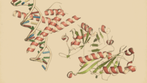
The Presence of α-syn
α-synuclein (α-syn), a protein closely associated with PD, isn't confined to the central nervous system (CNS); it also resides in the colon, the neurons of the enteric nervous system (ENS), and the vagus nerve A Trusted Source
Braak H, de Vos RA, Bohl J, Del Tredici K. Gastric Alpha-Synuclein Immunoreactive Inclusions in Meissner's and Auerbach's Plexuses in Cases Staged for Parkinson's Disease-Related Brain Pathology. Neurosci Lett (2006) 396(1):67–72. doi: 10.1016/j.neulet.2005.11.012 PubMed AbstractCrossRef Full TextGoogle Scholar .
This intriguing finding is mirrored in animal models of PD, where aggregates of α-syn have been detected in the GI tract A Trusted Source
Hallett PJ, McLean JR, Kartunen A, Langston JW, Isacson O. Alpha-Synuclein Overexpressing Transgenic Mice Show Internal Organ Pathology and Autonomic Deficits. Neurobiol Dis (2012) 47(2):258–67. doi: 10.1016/j.nbd.2012.04.009 PubMed AbstractCrossRef Full TextGoogle Scholar .
This discovery has led researchers to propose that PD might originate in the gut and subsequently spread to the CNS through the vagus nerve.
This theory gains support from animal studies revealing that externally introduced α-syn into the gut wall can journey to the brain via the vagus nerve,
covering a distance estimated at 5–10 mm per day in rats A Trusted Source
Holmqvist S, Chutna O, Bousset L, Aldrin-Kirk P, Li W, Bjorklund T, et al. Direct Evidence of Parkinson Pathology Spread From the Gastrointestinal Tract to the Brain in Rats. Acta Neuropathol (2014) 128(6):805–20. doi: 10.1007/s00401-014-1343-6 PubMed AbstractCrossRef Full TextGoogle Scholar ).
Additionally, a patient who underwent a surgical truncal vagotomy showed a reduced risk of developing PD later in life A Trusted Source
Svensson E, Horvath-Puho E, Thomsen RW, Djurhuus JC, Pedersen L, Borghammer P, et al. Vagotomy and Subsequent Risk of Parkinson's Disease. Ann Neurol (2015) 78(4):522–9. doi: 10.1002/ana.24448 PubMed AbstractCrossRef Full TextGoogle Scholar A Trusted Source
Hui KY, Fernandez-Hernandez H, Hu J, Schaffner A, Pankratz N, Hsu NY, et al. Functional Variants in the LRRK2 Gene Confer Shared Effects on Risk for Crohn's Disease and Parkinson's Disease. Sci Transl Med (2018) 10(423). doi: 10.1126/scitranslmed.aai7795 PubMed AbstractCrossRef Full TextGoogle Scholar .
Auto-Immune Reactions and PD
The exact cause and role of these gut disruptions in PD remain unclear, but the presence of α-syn in the ENS alone is sufficient to induce irregular colonic motility in the gastrointestinal tract,
which aligns with the severity of motor impairment observed in some animal models A Trusted Source
Paumier KL, Luk KC, Manfredsson FP, Kanaan NM, Lipton JW, Collier TJ, et al. Intrastriatal Injection of Pre-Formed Mouse Alpha-Synuclein Fibrils Into Rats Triggers Alpha-Synuclein Pathology and Bilateral Nigrostriatal Degeneration. Neurobiol Dis (2015) 82:185–99. doi: 10.1016/j.nbd.2015.06.003 PubMed AbstractCrossRef Full TextGoogle Scholar .
PD patients experiencing constipation exhibit increased infiltration of CD4+ T cells into the colonic mucosa, along with elevated circulating TH17 and Treg cells A Trusted Source
Chen Y, Yu M, Liu X, Qu H, Chen Q, Qian W, et al. Clinical Characteristics and Peripheral T Cell Subsets in Parkinson's Disease Patients With Constipation. Int J Clin Exp Pathol (2015) 8(3):2495–504. PubMed AbstractGoogle Scholar .
Furthermore, mice lacking PINK1 or Parkin genes, when exposed to bacterial intestinal infections, demonstrate increased permeability of the blood-brain barrier (BBB).
This facilitates the entry of pro-inflammatory cytotoxic CD8 T cells into the central nervous system (CNS).
These immune cells target the host's mitochondrial antigens, potentially leading to the elimination of dopaminergic neurons in the striatum and subsequent motor impairments A Trusted Source
Matheoud D, Cannon T, Voisin A, Penttinen AM, Ramet L, Fahmy AM, et al. Intestinal Infection Triggers Parkinson's Disease-Like Symptoms in Pink1(-/-) Mice. Nature (2019) 571(7766):565–9. doi: 10.1038/s41586-019-1405-y PubMed AbstractCrossRef Full TextGoogle Scholar ).
Inflammatory Markers
Histological data indicates increased immunoreactivity of the astrocytic marker GFAP in the colon of PD patients, along with elevated levels of inflammatory mediators such as TNF-α, IFN-γ, IL-6, and IL-1β A Trusted Source
Devos D, Lebouvier T, Lardeux B, Biraud M, Rouaud T, Pouclet H, et al. Colonic Inflammation in Parkinson's Disease. Neurobiol Dis (2013) 50:42–8. doi: 10.1016/j.nbd.2012.09.007 PubMed AbstractCrossRef Full TextGoogle Scholar .
Notably, these mediators are elevated in the early stages of the disease and are inversely correlated with disease duration.
Since both enteric and central glial cells respond to IL-6 and IL-1β, their upregulation could influence pain transmission in both the gut and the CNS A Trusted Source
Morales-Soto W, Gulbransen BD. Enteric Glia: A New Player in Abdominal Pain. Cell Mol Gastroenterol Hepatol (2019) 7(2):433–45. doi: 10.1016/j.jcmgh.2018.11.005 PubMed AbstractCrossRef Full TextGoogle Scholar .
Dysbiosis in PD-Induced Pain
Gut Barrier Permeability and PD
Gut dysbiosis might play a crucial role in the progression of PD by influencing the permeability of both the blood-gut barrier and the blood-brain barrier (BBB).
This can facilitate the transport of α-syn (alpha-synuclein), a protein associated with PD, from the gut to the brain.
Additionally, gut dysbiosis can activate sensory neurons, contributing to pain hypersensitivity.
These effects may occur directly or indirectly through the release of cytokines by immune cells A Trusted Source
Qi J, Buzas K, Fan H, Cohen JI, Wang K, Mont E, et al. Painful Pathways Induced by TLR Stimulation of Dorsal Root Ganglion Neurons. J Immunol (2011) 186(11):6417–26. doi: 10.4049/jimmunol.1001241PubMed AbstractCrossRef Full TextGoogle Scholar A Trusted Source
Liu T, Gao YJ, Ji RR. Emerging Role of Toll-Like Receptors in the Control of Pain and Itch. Neurosci Bull (2012) 28(2):131–44. doi: 10.1007/s12264-012-1219-5 PubMed AbstractCrossRef Full TextGoogle Scholar.
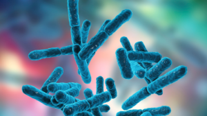
Characteristics of Dysbiosis
Dysbiosis in PD is characterized by increased levels of Enterobacteriaceae, Akkermansia spp., Catabacter spp., and Akkermansiaceae, alongside decreased levels of Roseburia spp., Faecalibacterium spp., and Lachnospiraceae A Trusted Source
Nishiwaki H, Ito M, Ishida T, Hamaguchi T, Maeda T, Kashihara K, et al. Meta-Analysis of Gut Dysbiosis in Parkinson's Disease. Mov Disord (2020) 35(9):1626–35. doi: 10.1002/mds.28119 PubMed AbstractCrossRef Full TextGoogle Scholar A Trusted Source
Lin CH, Chen CC, Chiang HL, Liou JM, Chang CM, Lu TP, et al. Altered Gut Microbiota and Inflammatory Cytokine Responses in Patients With Parkinson's Disease. J Neuroinflamm (2019) 16(1):129. doi: 10.1186/s12974-019-1528-y CrossRef Full TextGoogle Scholar.
Notably, Roseburia spp. and Faecalibacterium spp. are known for their anti-inflammatory properties and their ability to promote the release of anti-inflammatory cytokines A Trusted Source
Zhu J, Paul WE. CD4 T Cells: Fates, Functions, and Faults. Blood (2008) 112(5):1557–69. doi: 10.1182/blood-2008-05-078154 PubMed AbstractCrossRef Full TextGoogle Scholar A Trusted Source
Shen Z, Zhu C, Quan Y, Yang J, Yuan W, Yang Z, et al. Insights Into Roseburia Intestinalis Which Alleviates Experimental Colitis Pathology by Inducing Anti-Inflammatory Responses. J Gastroenterol Hepatol (2018) 33(10):1751–60. doi: 10.1111/jgh.14144 PubMed AbstractCrossRef Full TextGoogle Scholar.
Role of Roseburia spp. and Faecalibacterium spp.
Reduced levels of Roseburia spp. and Faecalibacterium spp. may exacerbate TH17 (T-helper 17) activity and limit the action of Treg (regulatory T) cells, potentially contributing to PD-induced pain A Trusted Source
Clos-Garcia M, Andres-Marin N, Fernandez-Eulate G, Abecia L, Lavin JL, van Liempd S, et al. Gut Microbiome and Serum Metabolome Analyses Identify Molecular Biomarkers and Altered Glutamate Metabolism in Fibromyalgia. EBioMedicine (2019) 46:499–511. doi: 10.1016/j.ebiom.2019.07.031 PubMed AbstractCrossRef Full TextGoogle Scholar A Trusted Source
Dekker Nitert M, Mousa A, Barrett HL, Naderpoor N, de Courten B. Altered Gut Microbiota Composition Is Associated With Back Pain in Overweight and Obese Individuals. Front Endocrinol (Lausanne) (2020) 11:605. doi: 10.3389/fendo.2020.00605 PubMed AbstractCrossRef Full TextGoogle Scholar.
Enterobacteriaceae and PD Symptoms
Higher levels of Enterobacteriaceae in the stools of PD patients are associated with motor symptoms and are linked to increased levels of lipopolysaccharide (LPS) and α-syn fibril formation A Trusted Source
Lin CH, Chen CC, Chiang HL, Liou JM, Chang CM, Lu TP, et al. Altered Gut Microbiota and Inflammatory Cytokine Responses in Patients With Parkinson's Disease. J Neuroinflamm (2019) 16(1):129. doi: 10.1186/s12974-019-1528-y CrossRef Full TextGoogle Scholar A Trusted Source
Bhattacharyya D, Mohite GM, Krishnamoorthy J, Gayen N, Mehra S, Navalkar A, et al. Lipopolysaccharide From Gut Microbiota Modulates Alpha-Synuclein Aggregation and Alters Its Biological Function. ACS Chem Neurosci (2019) 10(5):2229–36. doi: 10.1021/acschemneuro.8b00733 PubMed AbstractCrossRef Full TextGoogle Scholar.
While the direct link between Enterobacteriaceae and PD-induced pain is not established,
LPS is known to activate nociceptor neurons, possibly leading to heightened sensitivity in gut-innervating nociceptors A Trusted Source
Diogenes A, Ferraz CC, Akopian AN, Henry MA, Hargreaves KM. LPS Sensitizes TRPV1 via Activation of TLR4 in Trigeminal Sensory Neurons. J Dent Res (2011) 90(6):759–64. doi: 10.1177/0022034511400225 PubMed AbstractCrossRef Full TextGoogle Scholar A Trusted Source
Boonen B, Alpizar YA, Sanchez A, Lopez-Requena A, Voets T, Talavera K. Differential Effects of Lipopolysaccharide on Mouse Sensory TRP Channels. Cell Calcium (2018) 73:72–81. doi: 10.1016/j.ceca.2018.04.004 PubMed AbstractCrossRef Full TextGoogle Scholar.
Short-Chain Fatty Acids (SCFAs) and Pain
Bacteria in the gut produce SCFAs, including acetate, propionate, butyrate, and valeric acid.
These SCFAs can influence pain by driving microglia polarization, the release of inflammatory cytokines, and microglia activation A Trusted Source
Zhou F, Wang X, Han B, Tang X, Liu R, Ji Q, et al. Short-Chain Fatty Acids Contribute to Neuropathic Pain via Regulating Microglia Activation and Polarization. Mol Pain (2021) 17:1744806921996520. doi: 10.1177/1744806921996520 PubMed AbstractCrossRef Full TextGoogle Scholar A Trusted Source
Sampson TR, Debelius JW, Thron T, Janssen S, Shastri GG, Ilhan ZE, et al. Gut Microbiota Regulate Motor Deficits and Neuroinflammation in a Model of Parkinson's Disease. Cell (2016) 167(6):1469–80.e12. doi: 10.1016/j.cell.2016.11.018 PubMed AbstractCrossRef Full TextGoogle Scholar.
Additionally, SCFAs can affect the immune response by inhibiting Treg cells, increasing T cell density, and modulating leukocyte activity A Trusted Source
Smith PM, Howitt MR, Panikov N, Michaud M, Gallini CA, Bohlooly YM, et al. The Microbial Metabolites, Short-Chain Fatty Acids, Regulate Colonic Treg Cell Homeostasis. Science (2013) 341(6145):569–73. doi: 10.1126/science.1241165 PubMed AbstractCrossRef Full TextGoogle Scholar A Trusted Source
Vinolo MA, Rodrigues HG, Nachbar RT, Curi R. Regulation of Inflammation by Short Chain Fatty Acids. Nutrients (2011) 3(10):858–76. doi: 10.3390/nu3100858 PubMed AbstractCrossRef Full TextGoogle Scholar.
Sodium butyrate, an HDAC inhibitor, has been shown to reduce pain, suggesting a potential role of gut SCFAs in promoting sensory hypersensitivity A Trusted Source
Kukkar A, Singh N, Jaggi AS. Attenuation of Neuropathic Pain by Sodium Butyrate in an Experimental Model of Chronic Constriction Injury in Rats. J Formos Med Assoc (2014) 113(12):921–8. doi: 10.1016/j.jfma.2013.05.013 PubMed AbstractCrossRef Full TextGoogle Scholar.
Fecal Microbiota Transplantation (FMT) and PD: FMT has shown promise in restoring a healthy microbiome, decreasing SCFA levels,
alleviating physical impairment, and reducing neuroinflammation in PD animal models A Trusted Source
Sun MF, Zhu YL, Zhou ZL, Jia XB, Xu YD, Yang Q, et al. Neuroprotective Effects of Fecal Microbiota Transplantation on MPTP-Induced Parkinson's Disease Mice: Gut Microbiota, Glial Reaction and TLR4/TNF-Alpha Signaling Pathway. Brain Behav Immun (2018) 70:48–60. doi: 10.1016/j.bbi.2018.02.005 PubMed AbstractCrossRef Full TextGoogle Scholar.
In PD patients, FMT has led to improvements in constipation and certain motor and non-motor symptoms, as well as enhanced mental well-being A Trusted Source
Vendrik KEW, Ooijevaar RE, de Jong PRC, Laman JD, van Oosten BW, van Hilten JJ, et al. Fecal Microbiota Transplantation in Neurological Disorders. Front Cell Infect Microbiol (2020) 10:98. doi: 10.3389/fcimb.2020.00098 PubMed AbstractCrossRef Full TextGoogle Scholar A Trusted Source
Huang H, Xu H, Luo Q, He J, Li M, Chen H, et al. Fecal Microbiota Transplantation to Treat Parkinson's Disease With Constipation: A Case Report. Med (Baltimore) (2019) 98(26):e16163. doi: 10.1097/MD.0000000000016163 CrossRef Full TextGoogle Scholar.
There is growing interest in exploring how FMT could potentially improve PD-induced pain and delay the onset of motor symptoms A Trusted Source
Roman P, Abalo R, Marco EM, Cardona D. Probiotics in Digestive, Emotional, and Pain-Related Disorders. Behav Pharmacol (2018) 29(2 and 3-Spec Issue):103–19. doi: 10.1097/FBP.0000000000000385 PubMed AbstractCrossRef Full TextGoogle Scholar A Trusted Source
Pusceddu MM, Gareau MG. Visceral Pain: Gut Microbiota, a New Hope? J BioMed Sci (2018) 25(1):73. doi: 10.1186/s12929-018-0476-7 PubMed AbstractCrossRef Full TextGoogle Scholar.
Review date not set.
How we reviewed this article:
Latest on: